
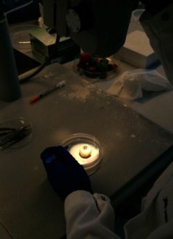
 This week we received some human eyeballs from which I was given tissue to culture cells. One of the lab’s MD, PhDs took the eyeballs apart. She started out removing muscle and parts of the optic nerve. Next she opened the eye balls and started separating the different parts. All of these different parts were then placed in tubes, which were then given to different people to extract and then culture cells from. I was given lens and stromal tissue. Once I was given the tissue the first part was cutting it into little pieces to make digestion – separating all the cells from the large clumps of tissue – easier. After I turned the tissue into little chunks, I added trypsin to the tissue and then placed the petri dishes back into the incubator and let it sit for a little for around 15-20 minutes. After that, I used the microscope to determine if the tissue needs more time to digest. After that, in order to inactivate the trypsin, I add media (2-3 times as much media as trypsin) in order to halt the digestion. The next step is to spin the solution down using one of the labs centrifuges. I transferred everything into a tube and then spun it down. Spinning down the tissue, cells, and media will result in the cells and tissue to gather at the bottom of the tube. After that, a vacuum is used to discard the supernatant, as only the cells are desired. The next step is to re-suspend the cells in media and transfer them into new wells. Then they go into the incubator and if everything goes according to plan, the cells start attaching immediately to the bottom of the plate.
Apart from starting new cell lines, I also did some DNA purification this week. At the lab we use kits (a couple of different companies provide DNA purification kits). The first couple of steps are designed to break open the cells, remove proteins, lipids, etc. These steps are followed by steps designed to precipitate and purify the DNA. The last step is to dissolve the DNA in a buffer solution. At this point the DNA is ready for analysis.
There are no comments published yet.