
This week I started the data and testing part of my project, which consisted of learning many new processes and collecting data to be used to figure out if the experiment has worked. This week my cells were grown enough, in their flasks, that I could take some of them out of their flasks and put them into their 96 well plates.
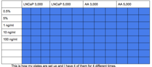
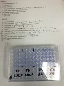
This is how my plates are set up. I have four of them for four different times.
To start the experiment I had to set up my layout for the plates. Each plate was a different time, so we had plates that we checked at 0 days, 2 days, 4 days, and 7 days. On each plate we had LNCaP (Caucasian prostate cancer) cells and African American prostate cancer cells and we had 5 different conditions with 3,000 cells per well in some and 5,000 cells per well in others. Our 5 different conditions were 0.5% serum in medium, 5% serum in medium, 1 ng/ml (nanogram per milliliter) of EGF, 10 ng/ml of EGF, and 100 ng/ml of EGF, and we did 3 trials for each condition so the data would be more reliable. EGF is Epidermal Growth Factor which is, at certain concentrations, supposed to make cells grow faster and better, so we were testing to figure out how the African American cells are affected differently, and our hypothesis was that the African American cells would grow better than the LNCaP cells. We made this hypothesis because all of the research we did stated that African American cancer cells are more aggressive and often grow much faster.
Special slide I put my solution with cells and trypan into to be counted.
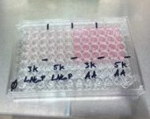
My Day zero, ninety-six well plate that I have marked on the cover so I know which wells have which cells.
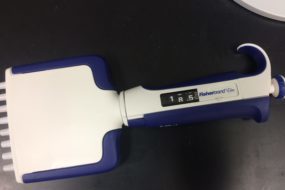
Multichannel pipet that I used to fill my ninety-six well plate.
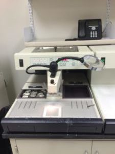
To transfer my cells from their flasks to the well plates like above, I had to first use this solution called trypsin to release the cells from the flask. Then after the cells are free from the flask I put them into a 15 ml tube. Next I take 20 ul (microliters) of the cells using a pipet and 20 ul of trypan (a type of blue dye) and mix them together. I then put 20 ul of the solution into each side of a special slide that is used for counting cells, then I insert the slide into an automated cell counter which then gives me the concentration of my cells per milliliter, so I can then calculate how many milliliters I need to get 3,000 cells per well and 5,000 cells per well. After my calculations I then put the needed amount of cells in each well using a multichannel pipet, which is a pipet that you can pipet multiple wells at once, and I put the correct medium or EGF into each well.
Automated cell counter that I put my slide in from above into to find the concentration of cells per milliliter.
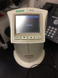
After that my plates are done until they have sat for the certain number of days that they are supposed to sit for. For my day zero plate I had to get it ready for cell counting for the control, so I take it through this process called crystal violet staining so the cells become stained so that another special machine can read how dark of a shade the stain is and by that, you can tell how much the cells change throughout the time period. Then I took my data and put it into graphs to show how much the cells have grown over the course of a few days. I have done this through day four and next week I will finish day seven and we will look at the data as a whole, and we will then see if our hypothesis was correct.
I also learned a new process of tissue embedding, which has nothing to do with my project but my mentors thought I would enjoy it, which I did, and they thought that since I wasn’t busy, while my cells grow, it would give me something to do. This process of tissue embedding is putting tissue specimen into wax and the cassette. Once the specimen is in wax it can then be used to make into slides so it can then be stained and examined.
Multichannel pipet that I used to fill my 96 well plate.
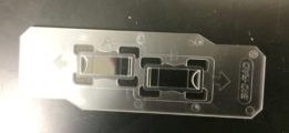
Machine used to embed specimen. The machine has a wax spout that dispenses hot wax and the flat part is all hot so the wax doesn’t harden except the little square in the middle is a cold spot so you can harden the wax a little to position the tissue specimen, and also there is a cold plate on the right of the wax machine that you put the cassette on when it is finished so it can harden quickly without being moved around too much.
This week has been the busiest week yet and I have probably learned the most this week out of any of the weeks. Next week I will be doing the same experiment but with used media from the cancerous stromal cells.
There are no comments published yet.