
![]() Hello everyone! I am Melaina Yender and this is my sixth and final week researching neuroscience and click chemistry at the Ye Lab at the Scripps Research Institute in San Diego, California. This has been such an incredible experience and I have taken away an immense amount of knowledge. I am very honored to have had the opportunity to work with Cailynn Wang and Zhengyuan Pang and research neuroscience through click chemistry. I am very grateful to have had the opportunity to work with both of them and to have worked in the Ye Lab.
Hello everyone! I am Melaina Yender and this is my sixth and final week researching neuroscience and click chemistry at the Ye Lab at the Scripps Research Institute in San Diego, California. This has been such an incredible experience and I have taken away an immense amount of knowledge. I am very honored to have had the opportunity to work with Cailynn Wang and Zhengyuan Pang and research neuroscience through click chemistry. I am very grateful to have had the opportunity to work with both of them and to have worked in the Ye Lab.
Monday was very special because the lab went on a fishing trip in Point Loma and had the opportunity to fish for most of the day. It was a very beautiful boat ride and we saw multiple dolphin pods, one of which consisted of around 50 dolphins! We also saw many different animals including jellyfish, seals, and some starfish. We caught a lot of fish and I cooked mine for dinner on Wednesday. After fishing, a family friend took me surfing at Pacific Beach for a few hours and it was so much fun!
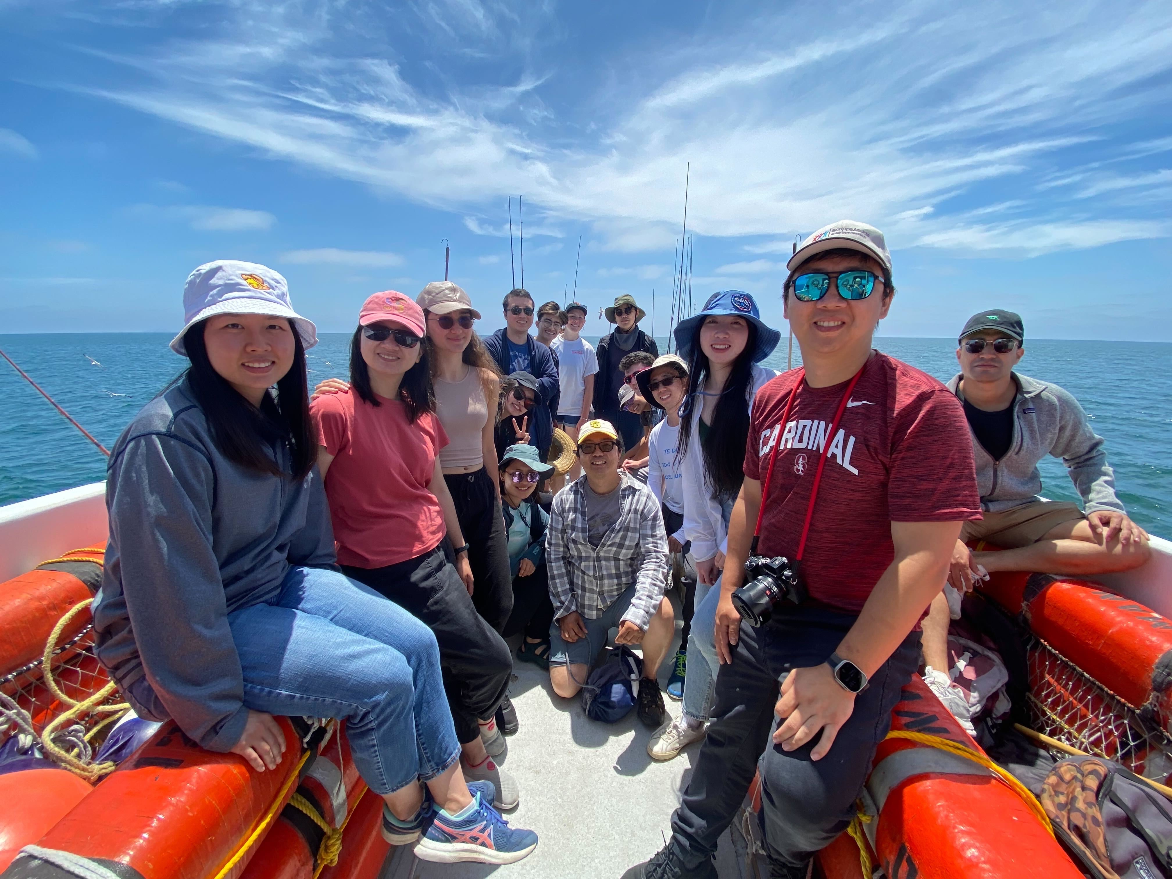
Ye Lab Fishing Trip at Point Loma!
To start the week, I washed my tissues that were degassed and polymerized over the weekend with PBST to get rid of the excess SDS. I then selected two different bregma from each of the samples, one anterior and one posterior, and incubated them in 200uL 1mM CuSO4 instead of the regular click incubation buffer. Since there was more intense labeling from the CuSO4 incubation from the experiment last week, I adopted this to my personal experiment. The following day I created the click reaction for the samples, washed them with PBST detergent, and then stained them with DAPI staining. Later in the week I mounted my slide with the samples and imaged them using confocal microscopy. The imaging came out great, and the new probe imaging was extraordinary and turned out better than expected. We also attempted the in vivo electrochem experiment again from the second week I was here. This was the process of directly electrifying the brain samples instead of creating lysates and extracting the membrane and cytosol fractions, and electrically activating the samples following that. This time the electrical activation was done on 500-micron slices instead of the full brain. This worked much better in comparison to the pilot experiment. In a couple of weeks, we will know if it actually worked to our expectations or not.
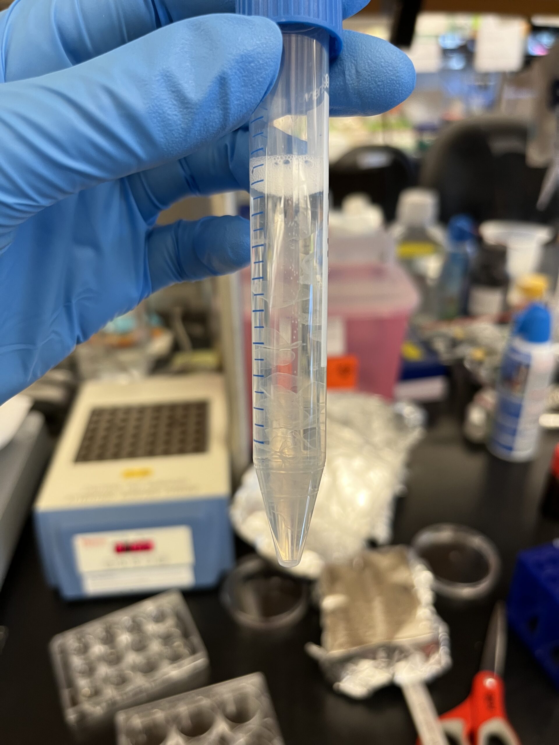
Washing cleared tissue samples in PBST
Two large discoveries were made at the beginning of the week on Tuesday. The incubation in CuSO4 was shown to have minimized the background reaction that the traditional incubation buffer created with the tissue. The side reactions with the samples caused the entirety of the tissues to become bright, which interfered with the labeling of the probe and often led to misinterpretation/confusion of where the probes were binding to in the brain tissue. This is major because many previous experiments have not gone to plan due to the excessive background reaction. The second discovery was that incubating the hemispheres in a high concentration of CuSO4 for a couple of days penetrated the cortex and occupied the proteins that the copper binds to. This is crucial because the previous incubation for volumetric samples did not effectively label the interior of the tissue which resulted in a lack of labeling inside, and only the cortex was labeled. This discovery allows for CATCH to be applied on whole organs rather than just thin slices of tissue, which can aid a lot in discoveries in the future. It has been shown that CATCH can expand beyond tissue slices and can be applied to entire mouse bodies. After this week, a lot of the original protocol was modified to include CuSO4 incubation for tissue samples.
On Wednesday, Ben wanted me to run an experiment with some vehicle samples that were not perfused thoroughly enough. He postulated that the CuSO4 incubation would minimize the labeling in the blood vessels and allows for the imaging of the blood vessels to not interfere with the signaling from the probe. I selected two slices from two different samples and incubated them in 1mM CuSO4 and in the traditional click incubation. I conducted the reaction for the samples and washed them using PBST detergent and followed with DAPI staining. From there I mounted my slide and imaged it using confocal microscopy. The CuSO4 incubation was proven to minimize the reaction in the blood vessels and resulted in clearer imaging. My mentors do not thoroughly understand why this is, but it was something that they noticed in the past week during all the CuSO4 incubation experiments. This means that the perfusion step of the procedure is not as necessary as previously believed, as extra CuSO4 incubation can take care of the imaging of the blood vessels.
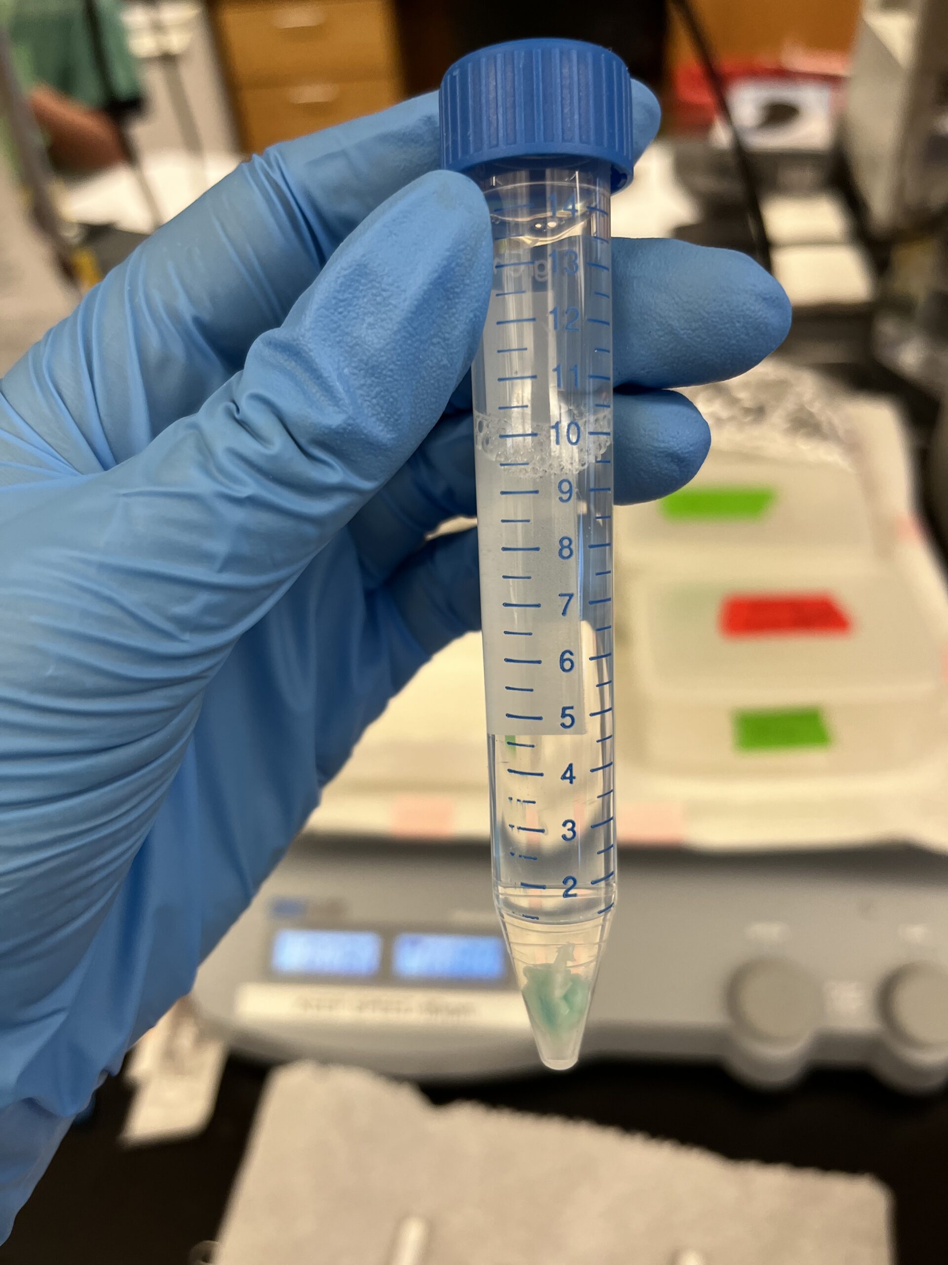
Hemisphere sample after CuSO4 incubation and multiple click reactions
Towards the end of the week, I also got the chance to run my own experiment with two pargyline-injected in vivo hemispheres that were already cleared a couple of months beforehand. I incubated them in 2 mM CuSO4 overnight and ran the click reaction the next day. The protocol for the hemispheres requires two days of incubation in CuSO4, however, I was limited on time this week. For one of the hemispheres, I ran the reaction only once and for the other hemisphere, I ran the reaction five times. The repetition of reactions for the samples is crucial to ensure that the maximum amount of probes is labeled through the entirety of the tissue. I then washed the samples with detergent and then cut the brains into 100-micron sections. Because I was short on time, I did not wash the samples enough times and the inside of the tissue was still a light blue from excess azide dye. Once I cut and imaged the samples, we noticed that the samples that underwent only one reaction were not labeled to the extent of the other hemisphere that underwent five reactions.
On my last day, my mentors took me out to lunch at this nice sushi place and we enjoyed a wonderful meal there. When we got back, Cailynn had bought a cake as a going-away surprise. I got to enjoy my last day with all the people in the lab which I enjoyed a lot. Before leaving, I went to the Scripps Pier with the other interns on our last night and watched a beautiful sunset. It was a great way to end my internship!
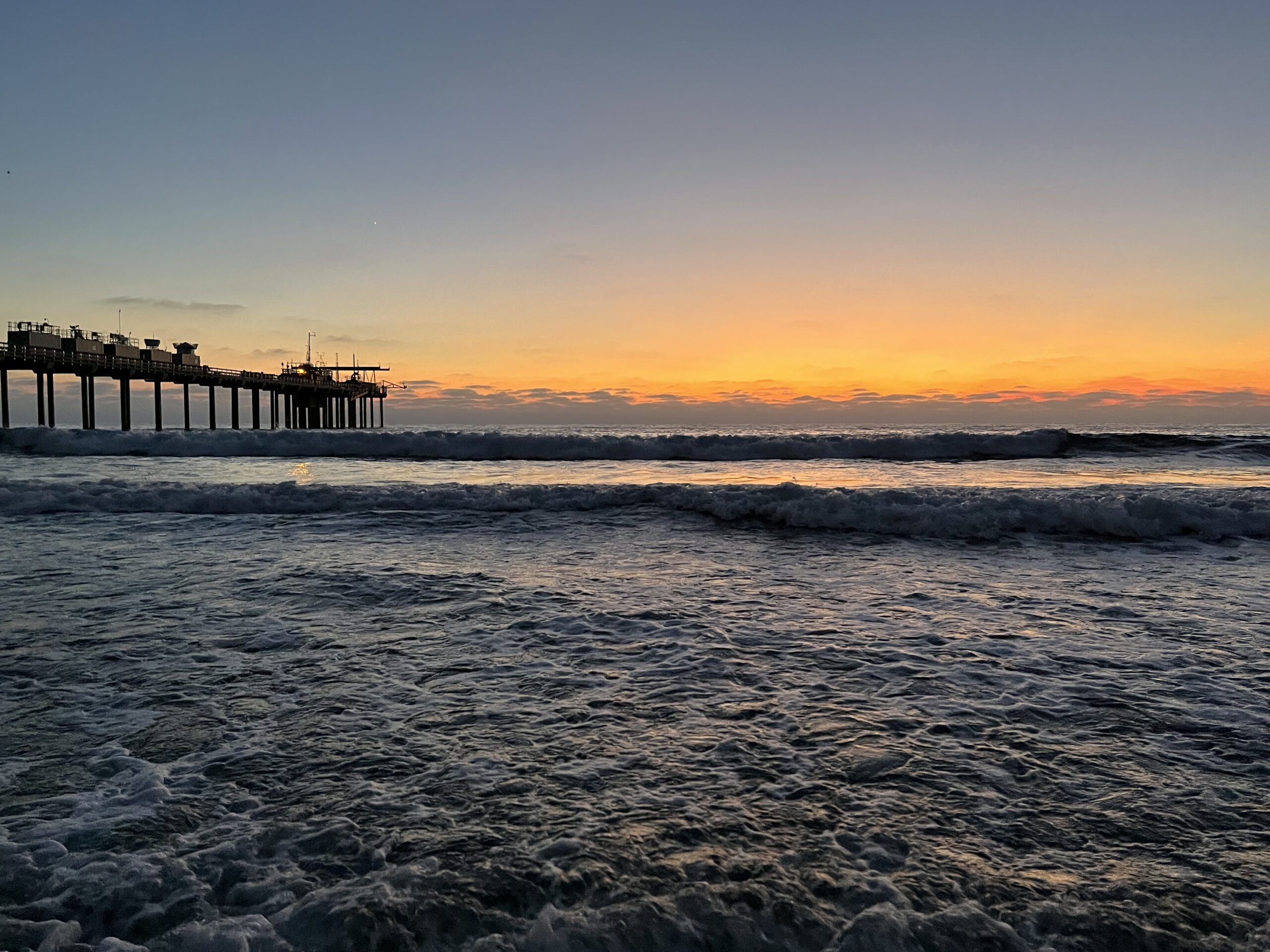
Last sunset at Scripps Pier
Again, I am so grateful for my internship and for everyone that I got to work with. I am also very thankful for Pinhead and Sarah Holbrooke for finding this extraordinary opportunity for me. This was truly the most enriching experience of my life, and I’m delighted to have been granted this opportunity!
There are no comments published yet.