
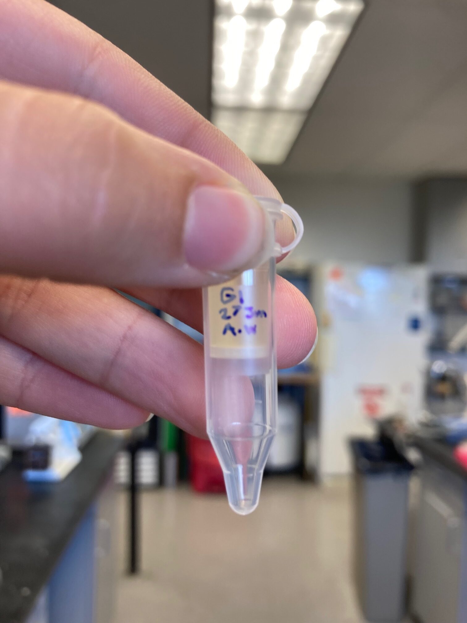
I am Aubrielle Weaver, spending my summer in San Diego, California, at San Diego State University (SDSU). For the next six weeks, I will be studying microbiology. My mentor is Gregory Burkeen, who is a Ph.D. student at SDSU. This is week 2 of my internship.
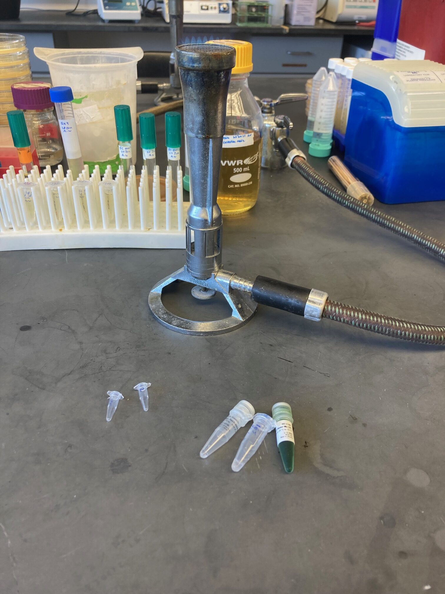
As soon as I came in on Monday, I went to check on my plates. I had tons of growth. My goat’s milk plate grew a lot, so it’s hard to distinguish different microbes. My cow’s milk got small colonies in one little area. The liquid culture plates got tiny little bacteria. And my other four plates had tons of growth. ¾ of the plates needed me to isolate the microbes further. One of the plates appears to have a fungus or yeast on it. The other two have bacteria. From each plate, I picked an isolated colony. I streaked them onto the plates I made Friday to continue to isolate them. I had also gotten growth on my cow’s milk plate, but it wasn’t distinct enough to isolate any microbes. So I streaked the little growth I had onto another plate. Greg also showed me how to place bacteria into a liquid media, which I put with my other plates in the incubator.

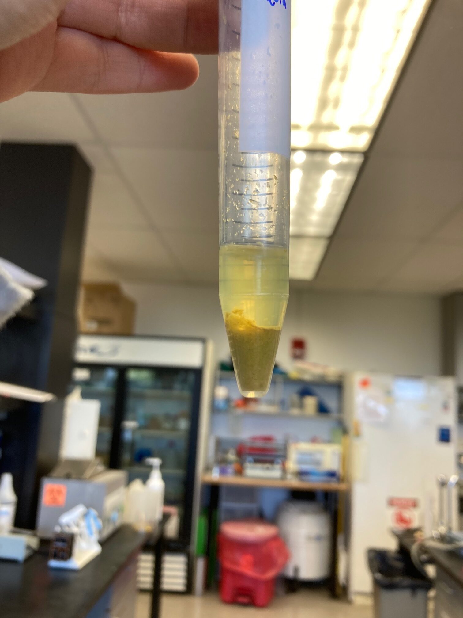
This is my liquid culture after a few days of growth. The foggy section is the bacteria.
We did the PCR and DNA electrophoresis to determine if our DNA was bacterial. From the plate with isolated bacteria, I took one colony and ran it through a thermocycler. It is the machine that carries out PCR. PCR is when hundreds of copies are made of a segment of the DNA in your sample. You add different Primers that correlate with the beginning part of the segment you want to be copied, and the enzyme taq polymerase will make the copies from that starting point. I wanted it to copy the V3 region of the 16s gene. If it copied this gene, that would confirm that my growth is bacteria. To know if it copied that section, I did the DNA Electrophoresis. A DNA Electrophoresis has a negative charge on one end of the device and a positive charge on the opposite. And in the center, you have a buffer, which protects the equipment and the DNA. And on this little platform, a square-shaped agar gel with some holes at the top side is placed in the buffer. The holes should be on the negative side of the device. The DNA is loaded into the holes, and you turn on the charge. After a while, the smaller fragments of DNA will move further down. You can’t see your results without putting the gel in a particular photo machine. This machine then scans your gel and, on the computer, pulls up an image. If no band appears, that means there wasn’t bacteria DNA. If a band appears, then it does have bacteria DNA.
These are the supplies needed to prepare your DNA sample.

This is an image of the PCR and Gel Electrophoresis results. The black streaks is the DNA that moved through the gel.
I observed Greg as these tests were done. And we did have a band appear, so we know it is bacteria. Once I have isolated the other microbes, we will send the DNA to be sequenced. Then we will know what bacteria they are.
The next day I came in and checked on the plates. The one that is supposed to have the suspected yeast/fungus had very tiny fungus spots and a lot of bacteria. Greg guessed that the fungus did not like the salts on the new media. He suggested that I streak it onto another milk agar plate to see if I could isolate it there. The second plate had no apparent growth; the third had little. And my cow bacteria looked close to isolated. In general, they look similar. I decided to incubate the plates longer and the milk agar plate streaked with the fungus.
I then made a subculture. I took 100 microliters of my bacteria from my liquid culture and five milliliters from my liquid media. I put it in the incubator to grow. In just two hours, you could already see some growth. After I finished up some research, I went out on campus in search of gross milky sources where I could potentially find Phages that would grow on my bacteria. I decided to start with campus trash cans in search of thrown-away coffee drinks. I first found what looked like a discarded mocha drink in a trashcan that smelled particularly bad. Then I found this mystery green smoothie drink that smelled horrible, so I took two samples from that. One from the more liquidy parts. And one from the chunky stuff floating in it. I also found a Starbucks cup with dried cream in it. Once I returned the samples to the lab, we centrifuged them to separate the liquid parts from the solid.
This is the green drink sample after being centrifuged.
The next part of the process is to filter the liquid layer of my samples in a 0.22 filter that will only let phages through and keep bacteria and other debris out. To do this, I measured some samples from the liquid layer. I put it into this tiny tube with two sections separated by the filter. I then centrifuged them to bring the stuff that could go through the filter down. The next part of the process is called an enrichment. While I waited for it to centrifuge, I equally divided my subculture into four tubes. I removed the filter layer when the samples were done in the centrifuge. I moved the liquid into their respective subcultures. We put the enrichments into the incubator to let phages reproduce with our isolated bacteria.
This is the filter tube before it was centrifuged. The sample is in the top portion on top of the filter and the bottom part is where the phages and sterile liquid will go.
The next day we retrieved my plates and enrichments. Our fungus/yeast grows best when it is attached to bacteria, or with the milk agar. The plate that I thought had no growth had tiny colonies. And then my third plate grew as expected but wasn’t entirely isolated yet.
After observing my plates, I then moved on to making another subculture. It was my first time doing something entirely on my own. I wasn’t perfect with my aseptic technique. The aseptic technique is how you limit contamination. Once I made the subculture, I moved my enrichments into new small tubes to be centrifuged. When that was done, a little pellet of bacteria was at the bottom of the tube. The rest of the liquid is called a supernatant. And I needed to transfer that supernatant into new tubes without getting any of the bacteria pellets. To do this, I used a micropipette. This allows me to grab a precise measurement. I was successful at moving only the liquid into the new tubes.
Before I could continue, I had to wait for my subculture to grow. So, in the meantime, I created some more media. This was my first time doing it by myself. I only needed a reminder on how to use the autoclave.
While I waited for my plates to dry, my subculture was ready to be used. I labeled one control and split the other into fours, each quadrant for a different sample. Greg placed 100 microliters of the subculture into the control plate. He added 1 milliliter of water and closed the plate. He swirled it to spread the liquid evenly, and then left it slightly open to dry. I did the same on the test plate. Once they were dry, I added 100 microliters of each sample to their assigned quadrant. Once dry, I put them into the incubator.
After grabbing my plates the next day, I observed that the bacteria grew evenly on both plates. However, there were no clear zones that would indicate viruses. This means I need to collect new samples. I’ll leave milk outside and use that. On my other plates, I had two new isolated bacteria—one from my cow kefir sample and one from my 2nd bacteria from the goat kefir. I ran a PCR and gel electrophoresis almost entirely on my own. I was successful in getting 16s amplification.
On Friday, I took my mystery fuzz and the last bacteria plate I needed to isolate from the incubator. The bacteria was finally isolated, so I ran a PCR. The fuzz can grow with minimal bacteria on a milk agar plate but needs bacteria to grow on the standard plate. I’m letting it grow more over the holiday weekend to see what happens. While waiting for the PCR and DNA gel electrophoresis to finish, I made liquid cultures for each of my isolated bacteria.
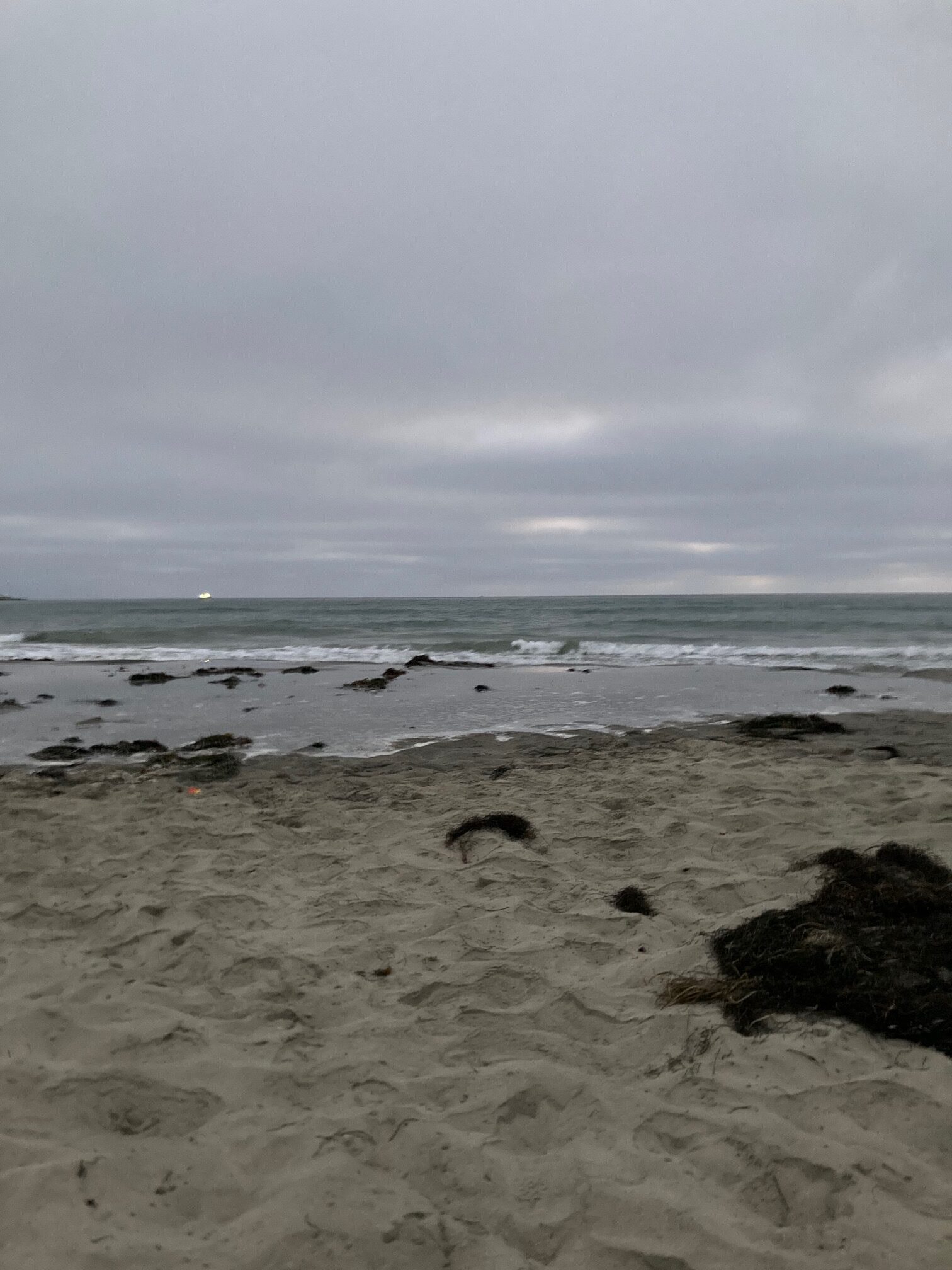
It was pretty cloudy during the weekend, but that didn’t stop us from going to the beach. Four more weeks left!
Seems as if you are learning a lot and enjoying the work keep on keeping on!
Aubrielle, sounds like you are having a blast. We are very proud of you. Keep up the great work!
Uncle Al and Aunt Dee