
Hello everyone! I am Melaina Yender and this is my second week studying neuroscience and click chemistry at the Ye Lab at the Scripps Research Institute in San Diego, California. I am involved in a process called clearing-assisted tissue click chemistry (CATCH) which is the process of clearing lipids from the cells in brain tissue and then tracing drug targets through the attachment of fluorescent tags on the drug molecules. This is crucial to understanding where certain drugs bind in the brain and understanding the specificity and how drugs will bind to other receptors and create adverse side effects. I explained this process more in-depth in my previous blog for week one.
This week was very busy as I was involved in several different processes and experiments. I was granted the opportunity to conduct my own experiment with the CATCH process. This gave me the chance to experience the entire CATCH process and grasp an understanding of how it works. I started at the beginning of the week by injecting four mice, two with the drug PF7845-yne and the other two with the vehicle as a control. An alkyne-modified version of PF7845-yne was used and is referred to as a probe. Alkyne-modified drugs are frequently used because they have the ability to bind with the fluorescent dye which can then be seen using confocal microscopy. The parental version of these drugs lacks this ability, so the use of probes is necessary. Thirty minutes following the injections I began to perfuse each mouse and harvest the brain. Perfusing is the process of pumping diluted phosphate-buffered saline (PBS) into the left ventricle of the heart and clearing the blood from the body through the removal of the right atrium. The goal is to remove all blood from the brain because the presence of blood affects the imaging of tissue slices. Then the same process is done with paraformaldehyde (PFA) to ‘fix’ the tissue. After PFA is pumped through the heart, the brain can be removed and stored in a cold room in PFA.
The following day I created an agarose gel to place the collected brain samples in and slice later on. The agarose gel maintains the position of the brain while it is being sliced and prevents anything from moving or going wrong. I had intended to slice the tissues that day, but there was not enough time due to a separate experiment that occupied the majority of the day. I was required to remove the tissues from the agarose because the water molecules will evaporate overnight and will essentially dry up and ruin the tissue. So the following day I recreated the agarose gel and was able to slice the tissues into 100-micron sections using a machine called a vibratome. After slicing I collected the tissue slices and stored them in a cold room in wells of PBS. They are now ready to be cleared this upcoming week and I am allowed to finish the rest of the CATCH process.
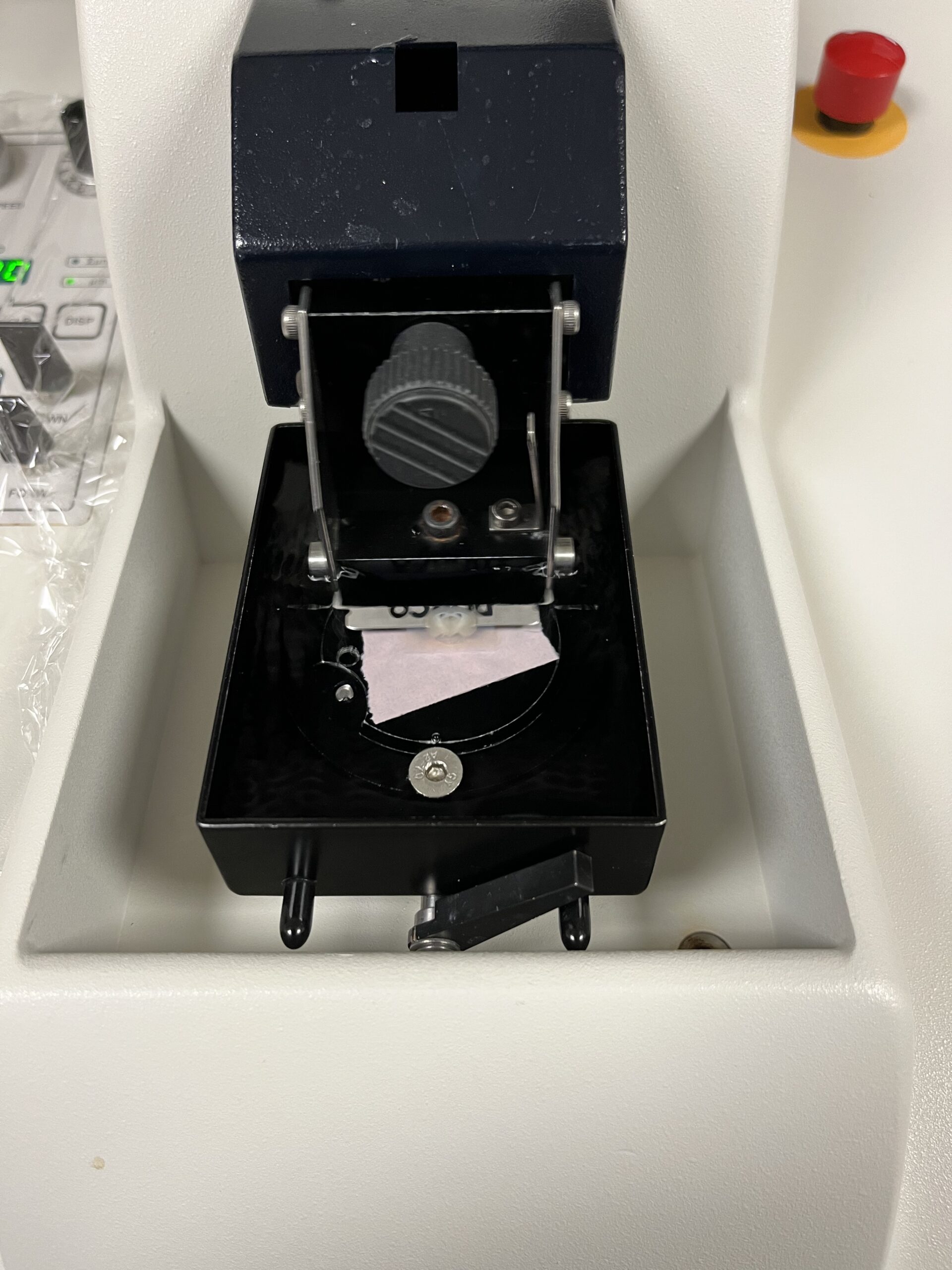
Vibratome machine
Aside from my experiment, I was involved with multiple other processes this week. My mentors had begun applying the CATCH process on lung samples from mice that were injected with Afatinib and Ibrutinib, which are pharmaceutical medications for lung cancer and leukemia respectively. Their goal is to see if this process can be done on full organs with the hopes of being able to image entire mouse bodies and observe drug targets across more than a 100-micron slice of tissue.
At the beginning of the week, the lung samples were degassed which removed oxygen molecules from the tissues and were then replaced with nitrogen. This process is crucial as oxygen would interfere with the polymerization of the hydrogel embedding and would bond with the polymers which would then quench the reaction. Following the polymerization process, the samples were embedded into agarose gel and could then be imaged using lightsheet microscopy. I had the opportunity to image one of the samples using the lightsheet microscope later in the week and observe the results.
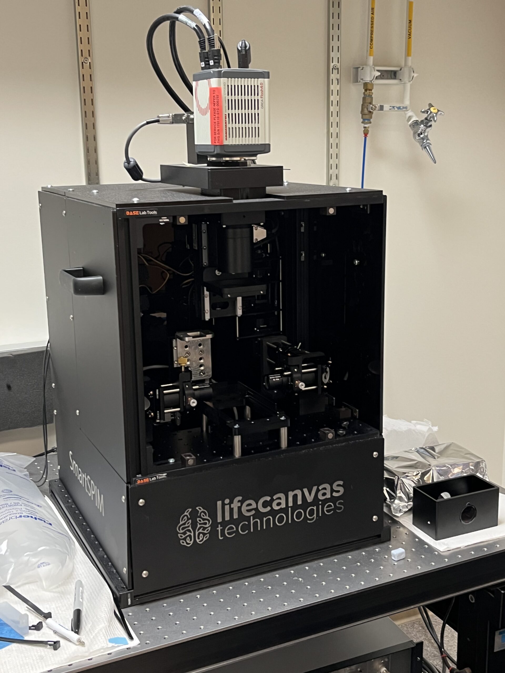
Lightsheet microscope used for imaging the lung samples
I was also involved in another experiment this week which was a second attempt with the electrochemistry experiment that had failed the previous week. The incompletion of the previous experiment was hypothesized to be due to an incorrect version of the drug which was unable to bind to the intended receptors. The mice were then injected with different alkyne modifications of the drug in hopes there will be potential binding to the receptors in the brain tissue. The procedure was modified and another technique of electrochemistry was applied to the lysates because the machine previously used for the whole brain tissue did not work as intended. Once the brains were harvested they were homogenized in cold PBS and placed in a centrifuge. The supernatant was then collected and prepared for electric activation. Half of the collected samples were electrically activated and the other half were used as a control. Half of the nonelectric vehicle group was diluted and used for in vitro treatment, which is the process of applying the drug directly to the tissue sample rather than injecting the drug in the mouse which is called in vivo treatment. Toward the end of the week, electrophoresis was used to separate the proteins from the samples using an electric current to pull the proteins through an agarose gel. The results supply knowledge on what proteins the
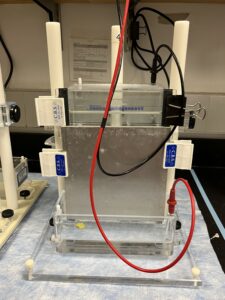
Electrophoresis process for the electrochem experiment
drug bonded to and if the drug had bonded to the tissue permanently and didn’t wear off.
This week was most definitely packed with experiments and I am so grateful to have the opportunity to be involved in these processes. In my spare time, I went to the beach on multiple occasions with the other interns I’m staying with. We all had Monday off from working and spent the day in downtown La Jolla. This week was absolutely wonderful and I am greatly looking forward to next week!
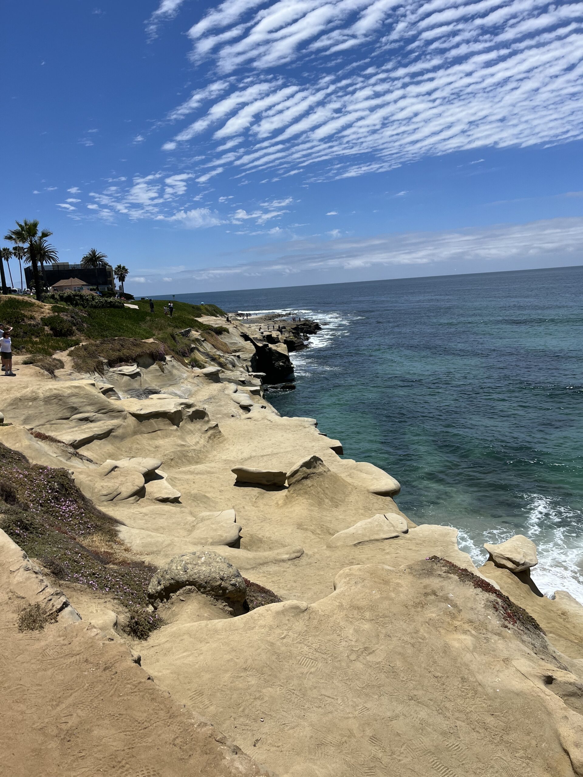
Picture of Whispering Sands beach in La Jolla
We sure enjoy reading you blog (I may not understand some of it ) so thankful you have this opportunity. God bless you. ❤️Nana
Melaine….this is very interesting. It will give you a great amount of insight knowing the complete process..what works and what does not work. Why to change tactics and which ones did not work and a reason why they didn’t work so as you move forward it will be easier to eliminate different processes. Learning how to read the data you collect, what you determine is very crucial so you can move forward the next week! Keep learning the new processes as it all builds on week 1. Hugs! We are proud of you!