
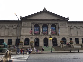
The Art Institute of Chicago from Michigan Avenue.
My fourth week at North Shore finally gave me some successful laboratory results. Since the past weeks of western blotting had produced no results, we were forced to make a third attempt at reading the protein expression levels inside our cell lines. However, due to a malfunctioning gel (into which we place the protein solution) on Monday, we were unable to continue. We discovered that our repeated attempts had completely used up the protein reserves we had made earlier, and we would have to wait until we could apply the treatment to a new group of cells, so we decided to move on to immunofluorescence testing.
On Tuesday, we started the test from scratch. Immunofluorescence is another common laboratory technique used for light microscopy in which specific antibodies bind to antigens (usually proteins in the cell) and are then given a fluorescent label that can be seen under a microscope, allowing researchers to have a visual of the intracellular processes and structures.
To be able to view the cell sample under a microscope, we first had to grow the cell cultures to a certain level of confluence and then place them onto a coverslip that will later be attached to a slide. After giving them ample time to attach to the coverslip in the medium, we administered the treatment of the oleic acid. Following the conclusion of the treatment period, we set about fixing the cells with formaldehyde, which essentially preserves the cells from decay and allows us to perform a test months after the initial treatment.
On Thursday, our cells were ready to be permeabilized, which creates holes in the cellular membrane that allows for the antibodies to enter the cell interior. After that, we applied two rounds of antibodies that target our specific protein and then applied a fluorescent stain called DAPI which allows us to detect the nucleus, giving us a better picture of the cell. Finally, we placed the coverslips onto the slides and sealed them off with a glue.
On Friday, we managed to take look at the cells under the fluorescence microscope inside a dark room. We were delighted to discover that the cells were in perfect condition and that we would be able to take detailed photographs that we could later analyze more. As these cells were cancerous, we also witnessed numerous instances of cell division (mitosis) being captured, which was truly an amazing sight. Next week, we will continue with the test for some of our other cell lines and hopefully will have good results.
Outside of the lab, I have also been able to meet with Telluride natives Christian Schumacher and Finn Bailis, both of whom are currently in the city. Furthermore, my host family has given me access to their membership for the Art Institute of Chicago, so I have been able to spend numerous hours viewing the works of the impressionists, modernists, expressionists, pointillists, surrealists, and romanticists.
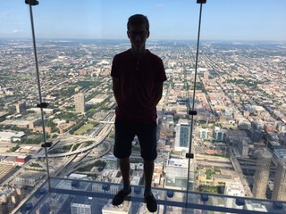
My silhouette in the Sears Tower glass balconies, more than a thousand feet above Chicago.
On Saturday, I also managed to look down on the entirety of the Chicago metroplex from atop the Sears Tower, which contains the highest floor in the western hemisphere. It was amazing to be able to view the jungle of concrete that is the city.
My time in Chicago has been great, and I look forward to continuing the next two weeks of my internship!
There are no comments published yet.