12 Jul Stella Coe- UCSD George Lab- Week 5
Hello everybody! Welcome back to my Pintern blog.
I can’t believe I only have one week left in the George Lab. This past week was absolutely jam-packed, and more than anything, it reminded me just how much I genuinely enjoy science. By the end of the week, I had assisted in 38 rat dissections—an experience that was equal parts fascinating, intense, and exhausting.
Monday 7/6
Monday was all about preparing the lab for an upcoming inspection. We worked our way through each room, making sure equipment was organized, supplies were stocked, and everything met the necessary standards.
I also received a new batch of catheters after they had been sterilized in an autoclave. An autoclave is essentially a giant pressure cooker used in laboratories. It uses extremely hot, pressurized steam to kill bacteria, viruses, fungi, and spores, ensuring that any surgical instruments are completely sterile before they are used again.

autoclave!
Later in the day, I analyzed another ELISA assay. After the plate reader measured the absorbance of each sample, I created a standard curve using samples with known nicotine concentrations. From there, I could determine the nicotine concentration in the unknown blood samples.
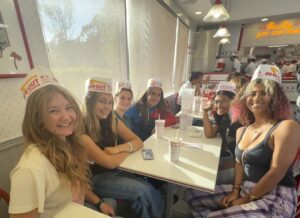
The day ended on a high note when AP scores were released. Thankfully, they were worth celebrating, so we made a trip to In-N-Out after work!!
Tuesday 7/7
Tuesday morning, I worked with Sonja at Skaggs preparing a sucrose solution for perfused brains. After a brain has been fixed in paraformaldehyde, it is transferred into sucrose, which slowly replaces the water in the tissue. This process cryoprotects the brain, preventing ice crystals from forming when it is frozen, so the tissue remains intact for future experiments.
Making sucrose with Sonja!
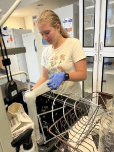
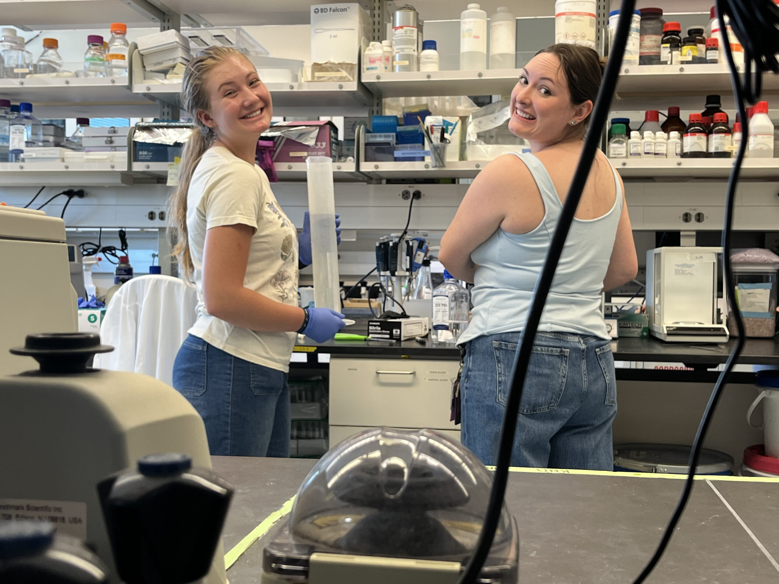
Keeping things clean!
While we were there, Sonja taught me how researchers map the brain using a brain atlas. Every location in the brain is referenced relative to a landmark called bregma, the point where several skull bones meet. Coordinates such as Bregma +0.72 mm (see photo) tell researchers exactly where a slice was taken. By comparing brain slices to the atlas, scientists can determine precisely where a virus expressing Cre recombinase infected neurons and which cells are expressing the fluorescent marker. Seeing the affected neurons glowing bright red made it much easier to appreciate how researchers confirm they targeted exactly the right brain region.
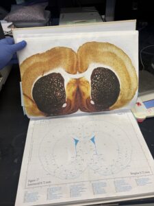 – Rat Brain Atlas
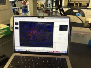
Red: affected neurons
The afternoon was much less exciting. I was scoring mouse behavior videos by counting how many times a mouse peeked over the edge of its enclosure. Unfortunately, I was so exhausted that I realized I could barely keep my eyes open. Since accurate data depends on careful observation, I told Selene that I couldn’t score the videos correctly. Without missing a beat, she simply asked, “Do you want to take a nap?” Fifteen minutes later, I was asleep on the lab desk. Honestly, it was exactly what I needed.
Wednesday 7/8
Wednesday was the day I had been looking forward to since the beginning of the internship… *dissections began*.
I’ll admit, the first few minutes were a little shocking. Watching the first rat be dissected was undeniably morbid. But very quickly, my perspective shifted. Every animal represented months of research, and every tissue sample would help answer an important scientific question.
Throughout the morning, I helped prepare syringes, labeled collection tubes, monitored the animals after CO₂ euthanasia to confirm they had passed, kept the paraformaldehyde reservoir full during perfusions, and organized each collected tissue sample. As organs were handed to me on a tray, I placed the brain, spleen, blood, and tail clip into their labeled tubes before immediately transferring them to dry ice.
The first time someone handed me a rat’s head on a tray was definitely startling. By the fifteenth one, though, it had simply become another step in the workflow.
One of the most interesting things was that there are two different methods for preserving brains, depending on what researchers plan to study.
Some brains were perfused. During perfusion, paraformaldehyde (PFA) is pumped through the circulatory system, replacing the blood and chemically cross-linking proteins throughout the tissue. This essentially preserves the brain exactly as it existed at that moment. After extraction, these brains remain in PFA before being transferred into the sucrose solution that I had prepared the previous day.
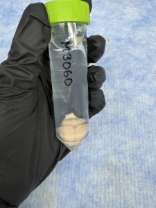
Perfused brain
Other brains were snap frozen. Instead of perfusing the animal, the brain is removed immediately after euthanasia and rapidly frozen in an extremely cold solution before being placed on dry ice. Because timing is critical, Sonja would call out, “Brain!” the instant one was removed, and I would drop whatever I was doing and sprint it over to the dry ice. It sounds dramatic, but preserving the tissue as quickly as possible helps maintain its molecular state for later analysis.
By noon, we had completed 19 dissections. The adrenaline that had carried me through the morning finally began to wear off.
Unfortunately, the day wasn’t over.
A new cohort of oxycodone rats had just arrived and needed to be scanned, identified, and placed into their cages in preparation for future Von Frey testing. My job was to create all of the cage cards so each animal could be easily identified throughout the study.
After lunch, I helped perform another round of Von Frey testing. Yes. More Von Frey testing. I thought last week was the last one, but I was sorely mistaken. Since these rats were younger and hadn’t yet become accustomed to handling, the process was definitely a bit more challenging.
And then… I got off work, and we all went to the Mormon Temple!! The temple was absolutely beautiful, and it was a wonderful group bonding activity.
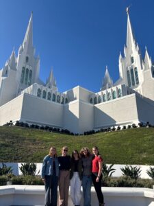
Thursday 7/9
Thursday looked similar to Wednesday as we completed the remaining dissections, bringing our total to 38 for the week.
The afternoon was just as busy. Lyena and I prepared clean cages for the new oxycodone cohort, scanned and labeled a new group of cocaine rats, assembled MRI-compatible catheters made with non-metallic components, weighed the EVSA rats, and cleaned their cages.
Somewhere in the middle of all of that, the long week finally caught up to us, and we were going a bit crazy. Our way to cope was singing Hamilton from memory while working. Apparently, that’s exactly what we needed.
When I got home that evening, I immediately collapsed into bed for a nap.
Friday 7/10
Kaila and I had a slightly later start Friday morning, so we took advantage of it by finally making it back to the gym after taking a short break during such a busy week.
Next Wednesday, we’ll be presenting our internship projects to the lab, so we spent part of the morning practicing our presentations with Sonja and making final improvements.
The rest of the day consisted of making more catheters, preparing rats for one last round of Von Frey testing, and—at long last—performing my *final Von Frey experiment*.
I have never been so happy to be done with a task.
While waiting for the EVSA rats to finish their session, Lyena and I watched Scrubs before weighing the animals. With that, another incredibly full week came to an end.
Saturday 7/11
Today was shopping at the mall day!! We had so so much fun at the UTC mall, and then that evening we went home and made Trang’s dumpling soup for family dinner! I am just soaking up these moments with the girls!!
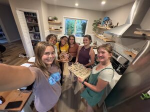
Dumplings!!
Sunday 7/12
Today is a beach day!
No Comments