

12 Jul Izzy Giroir – Neuroscience at Ye Lab, TSRI – Week 4
Welcome back to my blog! This week was my 4th week at the Ye Lab, and it was rather exciting as I got to see different pieces of the puzzle finally coming together! As a reminder, I’m working with researchers Kaili Xue and Leyao Shen, who are examining the metabolism of glucose during the excitation of neurons through processes such as learning and visual stimulation.
Monday (July 6th) –
I started my week off by attending a lab meeting about the findings of metabolism under different circumstances of stress. Factors such as pregnancy and the cold influence the neuron’s metabolism. For example, when cold, your body’s response is to naturally consume more food, which is used as fuel for different metabolic pathways. If I’m being honest though, I was only able to understand the beginning of the presentation, as described, but I still had fun looking at all the interesting graphs and listening to the feedback and questions provided by the lab as a whole. Just like the last lab meeting, I found that the environment was very collaborative. In the afternoon, Kaili and I went down to the sublevel to work with mice. Last week, we injected the mice with adeno-associated viral vectors (AAVs) that carried fluorescent proteins. These proteins make it so that whenever glucose is present, it is visually expressed by fluorescence. This expression is picked up and recorded by optic fibers that we also implanted into their brain last week. However, it takes about a week or two for the AAVs to fully diffuse, and so we did not use the mice that we operated on last week. Instead, we used mice that Kaili had operated on two weeks beforehand. These mice had double injections and insertions into their visual cortex, and so we did visual stimulation to pick up on the expression. The left side had been injected with an AAV that is expressed in inhibitory neurons (neurons that mute or block signals) and the right side had been injected with an AAV that is expressed in excitatory neurons (neurons that fire signals). To measure the consumption of glucose by these neurons, we hooked up wires to the optic fibers on the mice. These wires emit an excitation light at a specific wavelength (ex: 470 nm) to illuminate the target cells. In addition to this wavelength, a secondary control wavelength (ex: 405 nm) is used to monitor background noise. When glucose comes into the neurons, they emit fluorescence after being excited by the light, and the wires then carry the expression picked up by the fibers back to a computer, which translates the data into an active graph. First, however, we
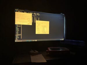
The computer that recorded the active data.
had to focus the excitation light concentration, which is done by looking at the overall expression of the injection site. When doing this, Kaili and I noticed that, on multiple mice, the right side injection site did not have much expression at all. Kaili told me this is likely because the area that she injected the AAV and the area she placed the insertion were slightly different, and were therefore not aligned. She decided to do a trial run of the visual stimulation (around 8 mins) before doing a full run (45 mins) to be sure that the data would still work regardless. The visual stimulation is done by placing tablets all around a plastic chamber.
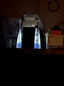
The visual stimulation paradigm (these mice are being trained to be iPad kids).
These tablets play a pattern of vertical black and white lines moving horizontally across the screen. You then put a covering inside of the chamber (along with the mouse), which blocks out the light from the screens completely. Then, you turn off all lights and start the data recording. Every 4 minutes, you raise the covering and allow the mouse to take in the screens for 15 seconds, then drop the covering back down. As the mouse is visually stimulated by the screens, the data records a dip in the glucose levels since the neurons are firing to process this information, and are therefore more rapidly consuming glucose. After the shorter trial run, Kaili decided that the data obtained would still be useful, so we continued on to an actual run-through, which took around 45 minutes. After the stimulation was over, Kaili and I went back upstairs to look over and analyze the data, which took up the rest of the day.
Tuesday (July 7th) –
In the morning, Leyao had me help her habituate a new batch of mice. Similar to my first week, I would place a mouse in a small chamber with a slit. I would also put a small glob of hydrogel (water in the form of jelly) in the chamber with it and give it 25 minutes to eat it and explore the chamber. After the 25 minutes was up, I would put it back into its cage and move on to the next mouse. I completed this process with 4 mice, and as I was by myself in the sublevel, I found the quiet very relaxing. I also took the opportunity to start a rough draft of my Common App while waiting (college applications are already haunting me). I would also like to note that I did not have a mouse make a run for it this time (iykyk). After lunch, Kaili and I completed another visual stimulation. While the mouse we used did have a double injection and insertion, the AAVs used were different. The left side once again measured the glucose concentration in inhibitory neurons, but the right side measured the overall neuron activity. This meant that the graph would show the glucose levels dropping as the neuron activity would spike. We also injected the mouse with 2-dg about 24 minutes into the stimulation. 2-dg (2-Deoxy-D-glucose) is a compound that mimics glucose and acts as a competitive inhibitor of the enzyme hexokinase. Simply, this means that the 2-dg will keep the glucose from being consumed during cellular respiration and therefore will create a buildup of glucose in the neurons. This specific stimulation was longer (about an hour and a half) so that we could fully record the rise, peak, and fall of the effects of the 2-dg. Being in a dark room, in complete silence, for an hour and a half is as tiring as it sounds. Luckily, I was the one raising and lowering the covering every four minutes, so there was no time for me to fall asleep. Kaili was not as lucky though . . .
Wednesday (July 8th) –
Similar to Tuesday, I habituated the mice in the morning and worked on my Common App rough draft in the morning. In the afternoon, though, I attended a subgroup meeting with Kaili, Leyao, and the other members in their cohort. Each of them briefly went over any important updates in their work as well as their works in progress and future plans. Afterward, the subgroup went to work on polishing their Powerpoints and so I read some articles on 2-dg to further familiarize myself with it. One of the articles presented findings on how 2-dg can inhibit the growth of cancerous cells. This is because cancer cells primarily utilize glycolysis (the specific step in cellular respiration that requires glucose) because it is the process that produces ATP the most rapidly. This is known as the Warburg effect. If you use 2-dg to inhibit glucose – and therefore glycolysis – the cancer cells cannot grow as exponentially. While this article didn’t pertain to the exact way I’m utilizing 2-dg, I found it pretty interesting!
Thursday (July 9th) –
Thursday morning, Kaili and I started the day off preparing to do some retro-orbital injection on mice with AAVs. These AAVs, however, target the mitochondria in the neurons instead of compounds such as glucose. We used PV-mito to target inhibitory neuron mitochondria and CamKII-mito to target excitatory neuron mitochondria. These AAVs also included an HA epitope tag. This tag is a short 10-amino-acid peptide sequence that is derived from the human influenza virus, and it attaches itself to the membrane of the mitochondria. Then, a couple weeks after the initial injection, you do a second injection that includes anti-HA antibodies. These antibodies find the HA tag and lock onto it. These injected antibodies are fused to small magnetic beads, which are used to separate the mitochondria from the rest of the cell when introduced to a magnetic field (this part is done when the mouse is dead and the brain has been taken out). On Thursday, we completed the initial injection on eight mice. To do this, we would anesthetize the mouse with isoflurane in an induction chamber. Then, you apply gentle pressure with your fingers to make the mouse’s eyeball protrude slightly. Using a syringe filled with the AAV solution, you insert the needle at a 45 degree angle into the corner of its eye with the bevel (opening of the needle) facing away from the eye. You do not insert the needle into the eyeball, but rather behind it. Once the needle hits the bone of the eye socket, you know it has entered the retro-orbital sinus – a complex network of veins located behind the eyeball in mice and other small mammals. You then inject the AAV solution, which is carried throughout the brain by the circulatory system. While this method of injection may seem cruel, it is actually considered one of the most – if not the most – humane way to deliver intravascular injections compared to alternate techniques in mice. After watching Kaili perform this on six of the mice, I got to give it a try on the remaining two! Later in the day, we perfused three mice (if you need a reminder on what perfusion is or how it is performed, refer to my second blog). These mice were three of the ones we operated on last week. Kaili needed to perfuse them to see if she got the concentrations of the injections right before she did the operation on a larger number of mice since this was the first time she had used some of these specific AAVs. Once again, after watching Kaili perform the perfusions on two of the mice, I was able to do the third one myself.
Friday (July 10th) –
Friday morning, I was upgraded to a new desk! The desk that I was at previously was being
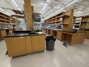
The empty lab I get all to myself!
shared between me and another undergraduate intern, and was in the area of heavy foot-traffic. Kaili was concerned I didn’t have enough space, so she showed me to the empty lab just down the hall. It’s been under construction, but the work on it has been paused for a bit. Now, I get an entire lab to myself, as well as get to sit next to a door ominously labeled “DON’T OPEN DOOR!!! DANGEROUS!”, which instantly makes it tempting. After getting settled, I helped Kaili prepare a new sucrose solution to put the brains we perfused yesterday in. Then, I transferred the brains from the tubes of PFA (that we had previously put them in the day before) into tubes of the sucrose solution. This is done to further stiffen/fix the brains so they are tougher and less likely to break when sectioning them with the cryostat. Afterwards, Kaili tried to book one of the surgery stations, but they were all filled for the day. I tried asking around the lab to see if anyone else needed help, but it seemed that it was a fairly slow day for everybody. To fill the time, I read papers on topics I had learned about during my time at the lab to further consolidate my knowledge, as well as to gain some new insight.
Saturday & Sunday (July 11th & 12th) –
Saturday, I went to the UTC mall with the other Pinterns here in San Diego, where Kaila and I saw Obsession while the others went shopping! It was nice to explore for a bit afterwards, and I was surprised that the mall was both giant and outdoors. On Sunday, my aunt and uncle took me to their friend’s house, who was kind enough to let us use her pool. I had fun relaxing in the water without having to worry about sand getting everywhere!
Once again, I would like to express how grateful I am for such an amazing opportunity! I’ve had so much fun this week and can’t wait to see you back here next week!!
Michelle Giroir
Posted at 15:40h, 20 JulyI love all the research reading you are getting the opportunity to dive into! Are you using CRISPR for the AAVs?