
28 Jun Izzy Giroir – Neuroscience at Ye Lab, TSRI – Week 2
With my second week already over, I’m excited to say that I’ve continued to learn so much! Though this week was a bit slower, I still got to accomplish many things.
Monday (June 22nd) –
To start my day off, I attended the weekly lab meeting where Xun Huang presented his findings on how the terminal structure of a neuron defined its function and role. Though most of the information went right over my head, I was still very interested in the parts that I was able to understand. It was also cool to see how collaborative the lab community is as they all pitched in to ask questions and answer them during the lab meeting. After the meeting was over, my mentors let me know that this week would likely be slower as they had a paper they needed to get published by a very quick deadline. As they didn’t have anything planned for me to help out with for the rest of the day, I took the opportunity to complete to rest of my training and onboarding tasks.
Tuesday (June 23rd) –
I started Tuesday by heading down to the sub level of the DNC where all the mice are located. This week, Leyao was working with a different set of mice and needed help habituating them again. This time, we used a maze to train them rather than a chamber with a slit. Another difference was that these mice had fibers implanted into their brain. These fibers are typically either inserted into the hippocampus or the visual cortex, and through the use of fiber photometry, they can measure the levels of calcium and glucose. This is vital for Leyao’s research as she is trying to understand the metabolism of glucose during learning processes and how that may be impacted by a mutant enzyme (PDH) within cellular respiration. However, these fibers also usually come with the downside of the mice being more sensitive and jumpy. I quickly learned this when one of the

This was the mouse that managed to jump out of the maze 🙁
mice managed to launch itself out of the maze and make a run for it. This was slightly terrifying since it was my first time handling the mice by myself, but I was able to catch it fairly quickly. After reporting this back to Leyao, she informed me that it wasn’t anything to worry about, which eased my concerns. After habituating the mice (by letting them roam around the maze for 15 minutes), I headed back up to the lab. I spent the second half of my day with Kaili, who helped me start a cell culture. We did this by preparing a high glucose cell culture media in a couple of petri dishes, thawing out some renal cancer cells that had been frozen in liquid nitrogen, adding the cells to the media, then placing the Petri dishes in an incubator to grow overnight.
Wednesday (June 24th) –
After coming into the lab, Kaili and I checked up on our cell cultures, which had expanded to the point where they took up the entire dish. We then dyed the culture with phosphorescent plasmids that had mixed into a buffer solution, then placed the cultures back into the incubator. This dye would make it possible for us to see the nucleus of the cells when excited with a certain wavelength under the microscope. Kaili and I then selected a handful of mice to perfuse (take out their brains). To do this process, you have to first euthanize the mouse by putting it in a chamber of isoflurane. You then cut open its chest till the heart is accessible and cut the right section of the heart as a secondary procedure to make sure the mouse is dead and not suffering. Next, you insert a needle attached to a tube into the left ventricle and pump the circulatory system with PBS (phosphate buffered saline) to clear the system of blood, then PFA
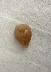
The brain that I took out myself.
(paraformaldehyde) to stiffen or “fix” its brain so it’s easier to remove. Finally, you cut into the skull, open it up, and remove the brain. We put the brains in a tube of PFA to further stiffen overnight. I found this process very interesting, and I even got to perfuse a mouse myself!
Thursday (June 25th) –
Once again, Kaili and I started our morning by checking on the cell cultures. This time, however, the cultures had mostly died off. Kaili told me that this was likely because the buffer had been contaminated with bacteria. We unfortunately just had to dump what was left of the cultures. In the afternoon, I went to the Intern Welcome Event that Scripps was hosting, where I ate lunch and met some other interns. Once I got back to the lab, I transferred the perfused brains from the tube of PFA to a tube of sucrose solution, which helps to further dehydrate the brain. I also cleaned some of the fiber implants that had been taken out of mice and were going to be reused. I finished my day by meeting up with another Pintern – Zoe Schiffer. It was nice to be able to talk with her and listen to her experience at Scripps!
Friday (June 26th) –
In the morning, my mentors were working on the paper again and most of everybody else in the lab was in a meeting, so there wasn’t much for me to help out with. Because of this, I researched how fiber photometry and confocal microscopes work, which I found very helpful in understanding why my mentors use the techniques they do. In the second half of the day,

The brain slices that I mounted onto a slide.
Kaili and I took the perfused brains out of the sucrose and prepared them for the cryostat by putting them into gel and freezing them over dry ice. We then took them over to the cryostat to be cut into very thin slices. Next, we put them into a buffer solution and mounted them onto slides to be looked at under the confocal next week.
Saturday & Sunday (June 27th & 28th) –
I spent most of Saturday hanging out with my Aunt and Uncle and playing board games, so it was a nice day of rest. Today, we went to the San Diego County Fair, which was fun because the fair that usually comes to Montrose is pretty small!
Overall, it’s been a very enjoyable week and I continue to look forward to the rest of the internship experience!!
Michelle Giroir
Posted at 15:24h, 20 JulySuch fascinating work you are doing. I am so thankful you have this opportunity!