19 Jul Izzy Giroir – Neuroscience at Ye Lab, TSRI – Week 5
My fifth week here at the Ye Lab has been very exciting, and I can’t believe my internship is already almost over! As a reminder, I’m working with researchers Kaili Xue and Leyao Shen, who are examining the metabolism of glucose during the excitation of neurons through processes such as learning and visual stimulation.
Monday (July 13th) –
I started my day on Monday by preparing the brains we had perfused last week for the cryostat. To do this, I took them out of the sucrose solution they were in (which helps to dehydrate them) and dried them off. Then, I put them in small plastic wells and embedded them within OCT – Optimal Cutting Temperature compound – which is a water-soluble gel (usually a blend of glycols and resins). We then put the well over dry ice in order to freeze the gel and the brain within. After I had prepared them, I placed them in the freezer to store them for when Kaili and I are ready to take them to the cryostat. After lunch, Kaili and I then headed down to the vivarium to do some surgeries. In total, we completed two operations, both of them being double injections and insertions into the CA1 region of the hippocampus (if you need a reminder of how these surgeries are done, refer to my blog from week three). Because the CA1 region of the hippocampus is very medial (close to the midline of the body), and the optic fiber cannulas are large, you cannot insert the cannulas perpendicular to the skull as usual. If we were to try to do so, we would not be able to place the 2nd cannula because it would bump into the first one. To fix this issue, we instead insert the cannulas at a 10 degree angle. The rest of the surgery, however is unchanged from the usual procedure.
Tuesday (July 14th) –
On Tuesday, we completed another two surgeries (double injections and insertions into the CA1 region). Typically, I do the beginning and end of the surgery by myself while Kaili does all of the locating and injecting in the middle. This was still the case for the first surgery we did. However, during the second surgery we did in the afternoon, Kaili let me do everything myself. While I was very excited, it was definitely a learning experience! Locating all of the points on the skull while still taking care to not break the glass needle was a lot more difficult than I thought it would be. In the end, I was able to complete the operation on my own, though it did take three hours (compared to the usual one and a half).
Wednesday (July 15th) –
In the morning, Kaili and I went down to the vivarium to prepare a room for the visual stimulation paradigm with the 2-dg injection. However, we realized that the room we needed to use was already booked for the day, and all of the surgery stations were as well. Because there were no hands-on experiments for us to do, I took the opportunity to read up on some more research papers. I think I ended up reading a total of three (all on varying topics of metabolic neuroscience), which took up a good chunk of time. Afterwards, I started working on my brag sheet in preparation for college applications and letters of recommendation.
Thursday (July 16th) –
On Thursday, Kaili and I completed three more surgeries, once again double injections and insertions into the CA1 region. During the time that Kaili worked on locating and injecting, however, I went back upstairs to help set up some new chambers. These chambers are the
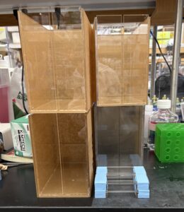
The new chambers that I assembled.
same type that I used to help Leyao with for her mouse training experiments – the ones with a small slit down the middle of the front panel. I set up a total of three – one during each surgery. Later in the afternoon, I joined Melaina (a former Pintern from Montrose) in the histology room where she was slicing some brains with the vibratome. Similar to the cryostat, the vibratome sections the brain into very thin slices so they can be imaged. Unlike the cryostat, the vibratome does not require the tissues to be frozen in OCT. The brains are instead put into agarose to fix them in place. They are then superglued to a mount, submerged under PBS, and locked into place. Then, a very sharp blade will cut through the

This is what a most of an entire brain looks like when sliced with a vibratome.
brain, leaving thin slices to float in the PBS. Once the blade cuts down to the area of interest (in this case it was the hippocampus and the posterior sections), the slices are removed and placed into a petri dish filled with additional PBS. In total, Melaina sectioned eight brains. Afterwards, Melaina took me out to dinner – where we both got tacos from a local Mexican restaurant – then very kindly drove me home. (Thank you, Melaina!!)
Friday (July 17th) –
On Friday, Kaili spent the day working on her PowerPoint, so I once again turned to reading papers! In the late morning, though, I did briefly accompany Melaina as she transferred the brains she had sectioned, and told me more about CATCH and click chemistry.
Saturday & Sunday (July 18th & 19th) –
Saturday, I went to Balboa Park with Ellie, Belle, and Zoe. We explored the many beautiful gardens, visited two of the museums, and played around in the interactive entrance zone of the
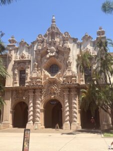
Some of the beautiful architecture at Balboa Park.
Fleet Science Center! Today, I went with my aunt to watch The Odyssey (which, if you haven’t watched it yet, I highly recommend).
This has been such a wonderful opportunity so far, and I can’t wait to see what my 6th and final week has in store!!
Glenn Giroir
Posted at 21:38h, 20 JulyYou are doing such incredible things. I’m so excited for you and proud of you!