
05 Jul Izzy Giroir – Neuroscience at Ye Lab, TSRI – Week 3
My third week at Scripps has been, while somewhat repetitive, very enjoyable! As a reminder, I’m working at the Ye Lab at the Scripps Research Institute. My mentors (Kaili Xue and Leyao Shen) are looking into the metabolism of glucose during learning processes, and how such metabolism can be altered by a mutated gene within cellular respiration. We worked on mouse surgeries throughout this entire week.
Monday (June 29th) –
On Monday, Kaili let me know that we would be working primarily on mouse surgeries. During these surgeries, we would be implanting optic fibers into the mice’s brains that would allow us to monitor active calcium and glucose levels. This is specifically done by anesthetizing the mouse
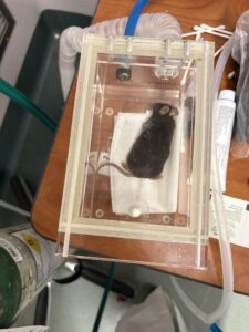
The induction chamber we used to anesthetize the mice.
using isoflurane, initially in an induction chamber. Then, you would hook the mouse’s front teeth into a hole in a metal plate (to keep their head stable) and attach a nose cone to keep a continuous stream of isoflurane. You then place metal bars on both sides of the mouse’s head to further stabilize it. After that, you put gel over the mouse’s eyes to keep them from drying out. For whatever reason, mice rarely close their eyes when being anesthetized and it is important for us to keep their eyes healthy as we are trying to stimulate their visual cortex. Next, you need to remove the hair from their head. We do this by applying Nair (this was my first time
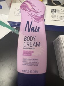 The bottle of Nair we used.
experiencing the awful smell of Nair) to their fur, then taking off the fur and washing their head with a saline solution and some q-tips. After the fur is removed, you cut off the skin around the skull until a decent circle is exposed and apply hydrogen peroxide to the skull to take off the membrane. The membrane is rather slippery and would cause issues when trying to attach the fibers to the skull, which is why we take it off. To further make the skull more adhesive, we also score it using a scalpel. Kaili would then use a microscope with an attachment to find the coordinates of the visual cortex. The attachment was hooked up to a machine with x,y, and z coordinates to give a precise location. Kaili would start by finding the bregma of the skull and zeroing out the x and y coordinates. She would then locate the lambda and zero out the z coordinate. Then, she would move the attachment to the recommended coordinates of the right visual cortex. She would then insert a glass needle into the attachment, which was filled with an adeno-associated viral vector, also known as an AAV. AAVs are viruses that don’t cause disease and are used them by removing the virus’ genes and replacing them with other materials. In our case, we replaced them with a fluorescent protein. This protein makes it so when our chosen molecule (glucose or calcium) binds to receptors in the neurons, it emits florescence that the optic fibers pick up on and record. To inject this AAV, Kaili would drill a hole in the skull at the coordinates she found, then insert the needle and set some parameters into a machine that would control the amount of virus injected within a time frame. We injected 300 microliters in three minutes, then waited an extra ten before we took the needle out to ensure that the virus had diffused. Then we started the process of attaching the optic fiber by switching out the needle for a optic fiber cannula, once
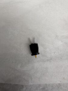
One type of optic fiber cannula we used.
again lowering it through the hole in the skull. Using dental cement, we secured the fiber to the skull, and to be extra cautious, we further secured it by painting on resin and curing it with a UV light. We did this surgery on three mice in total on Monday.
Tuesday (June 30th) –
On Tuesday, we did more surgeries. Once again we did a single AAV injection and fiber insertion into the visual cortex, but only on one mouse. For the other two mice we operated on, we did a double injection and insertion into the visual cortex (left and right side). This is because we wanted to monitor both glucose and calcium instead of just one or the other.
Wednesday (July 1st) –
We started our day with another surgery. Once again, it was a double injection and insertion into the visual cortex. After this first surgery, though, Kaili and I took a break to attend a meeting about a new microscope, specifically the two-photon microscope. From what I was able to understand, this microscope would allow the mice to freely move during experiments and the microscope would still be able to read and record high resolution images and data. This is crucial for certain experiments as the mice may act differently when they are not able to freely move. After the presentation, Kaili and I completed one more surgery. This time, instead of focusing on the visual cortex, we did a double injection and insertion into the hippocampus. This region of the brain (specifically the CA3 region of the hippocampus) is geared towards learning. Because of this, we will be testing these mice with learning stimulations rather than visual stimulations.
Thursday (July 2nd) –
Much like the days before, Kaili and I worked on some more surgeries. Today, we completed a total of three double injections and insertions into the CA3 region of the hippocampus.
Friday (July 3rd) –
Because the 4th of July lands on a weekend instead of a week day, we were given the 3rd off. I took the chance to meet up with the other Pinterns in San Diego. We all went to the beach, rode a roller coaster, and got boba together. While I may have gotten a little (a lot) sunburnt, it was nice to meet them all and have the opportunity to spend the day with them.
Saturday & Sunday (July 4th & 6th) –
For the 4th, I spent the day playing board games and watching movies with my aunt and uncle, then went out to watch the fireworks! Today (Sunday) we went to the beach again, and this time, I took care to reapply sunscreen frequently!!
Overall, while this week was a lot of the same thing, I found the surgeries very interesting, especially since I was able to do about 75% of the process myself! It was also great to be able to talk to Kaili and get to know more about her in the waiting time during the surgeries (which was a lot as each surgery took about 1 hour and 45 minutes). I can’t believe I’m already halfway done, and I’m so happy I’ve gotten to experience such a wonderful opportunity! See you next week!
No Comments